
Silent growths you need to know about
Let me share something that surprises most of my patients. Up to 70% of women develop uterine fibroids by age 50, yet very few understand what these growths actually are or how modern imaging can detect them. Many women live with symptoms for years without realising a simple scan could provide clear answers. Many women carry fibroids without ever knowing—they cause no trouble at all. But for others, these benign growths quietly disrupt daily life with heavy periods, pelvic pain, or pressure that just won’t go away.
Here’s the good news: today’s imaging technology gives us an incredibly clear view inside your body. We can spot fibroids early. We can map their exact size and location. And we can help your gynecologist choose the right treatment for you.
What exactly are uterine fibroids?
Think of a fibroid as a knot of muscle tissue that grows in or around your uterus. Doctors call them leiomyomas or myomas. Every single one of them is non‑cancerous. Having fibroids does not put you at risk for uterine cancer.
Fibroids come in different shapes and locations:
- Intramural fibroids grow within the uterine wall itself.
- Subserosal fibroids push outward from the uterus.
- Submucosal fibroids bulge into the uterine cavity.
Some women have one fibroid. Others develop several. Size varies enormously—some measure smaller than a pea, others grow as large as a melon. A study in the UAE documented the removal of 168 fibroids from a single woman’s uterus, highlighting just how extensive this condition can become.
A regional systematic review found that approximately 30.6% of women in the Middle East have uterine fibroids. That means nearly one in three women you know may carry these growths.
Could fibroids be causing your symptoms?
Many women dismiss their symptoms as “just part of being a woman.” Please don’t.
Fibroids can cause:
- Heavy or prolonged menstrual bleeding (sometimes with large clots)
- Pelvic pain or a feeling of pressure
- Frequent urination
- Pain during intercourse
- A visibly enlarged lower belly
- Difficulty conceiving or pregnancy complications
In the UAE, where many women delay childbearing into their 30s or 40s, fibroids often coincide with years when fertility is already more challenging, compounding both emotional and medical stress. Early detection matters enormously—not just for your comfort, but for your future family plans.
How we actually find fibroids with modern imaging
Gone are the days when discovering a fibroid meant guessing or waiting for severe symptoms. Today, we have a clear pathway to diagnosis, starting with the simplest and least invasive tools.
Ultrasound: your first window into the uterus
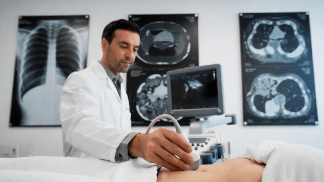
Ultrasound is always our starting point. A transvaginal ultrasound (TVS) gives us the best view of your uterus and ovaries. The procedure is quick, painless, and completely radiation‑free. It confirms whether fibroids are present and lets us count them and roughly measure their size.
On ultrasound, fibroids appear as well‑defined masses within the uterine wall. We can tell where each fibroid sits—inside the cavity, within the muscle, or pushing outward. That information alone often guides the next steps.
For many women, ultrasound provides all the answers they need. But for complex cases, we turn to more advanced technology.
MRI: the gold standard for fibroid mapping
Magnetic resonance imaging (MRI) offers something ultrasound cannot: exquisitely detailed pictures that show fibroids, blood vessels, and surrounding structures with remarkable clarity. We use MRI when:
- You have very large or multiple fibroids
- Your doctor needs to plan surgery or a procedure like uterine artery embolisation
- Ultrasound images are unclear or inconclusive
- We need to rule out other conditions like adenomyosis
MRI does not use radiation. The scan typically lasts 30–45 minutes. Some patients worry about claustrophobia, but modern machines offer wider bores, and we can provide music or even sedation if needed.
MRI can change the treatment plan in up to one‑fifth of patients. That is why advanced centres like Al Safwa Radiology offer both ultrasound and MRI—so women receive the precise information they deserve before making decisions.
Hysterosonography (saline infusion sonogram)
Sometimes a standard ultrasound does not clearly show the inside of your uterine cavity. That’s where hysterosonography comes in. We inject a small amount of sterile saline into your uterus through a tiny catheter. The fluid expands the cavity, allowing us to see fibroids that bulge inward (submucosal fibroids) with much greater detail.
The procedure takes only a few minutes and causes mild cramping at most. Hysterosonography has 92% sensitivity for polyps and 85% for fibroids, making it an exceptionally reliable tool.
Standardised reporting: the FIGO classification
You may wonder why imaging reports include numbers and types. Radiologists now follow the FIGO classification system—a universal language that describes exactly where each fibroid sits in relation to the endometrium and uterine wall. When your gynaecologist sees a “FIGO type 2 submucosal fibroid,” they instantly understand its precise location and can plan treatment accordingly. Consistency in reporting means no confusion and better outcomes for you.
From imaging to treatment: what happens next?
A radiology report does not just describe fibroids—it guides your entire treatment journey.
For mild or no symptoms
If your fibroids cause no trouble, we may simply monitor them with periodic ultrasounds. Many women never need intervention.
For symptom relief without major surgery
Medications like hormonal IUDs or birth control pills can reduce heavy bleeding, though they do not make fibroids disappear. These options work well for women who want to manage symptoms without procedures.
Uterine Artery Embolisation (UAE)
This minimally invasive, non‑surgical procedure has transformed fibroid care. An interventional radiologist guides a tiny catheter to the arteries feeding your fibroids and injects microscopic particles that block blood flow. Deprived of oxygen and nutrients, the fibroids gradually shrink. Studies show UAE improves symptoms in 85–90% of patients.
No large incisions. No removal of your uterus. Most women return to normal activities within one to two weeks.
Myomectomy
For women who wish to preserve fertility, surgeons can remove fibroids while leaving the uterus intact. This operation works well for certain fibroid types and locations.
Hysterectomy
Removing the uterus remains the only definitive cure for fibroids, but most women today choose less invasive options first.
Why choosing the right radiology centre in the UAE matters
Not all imaging centres provide the same quality. When you need fibroid evaluation, look for:
- Modern equipment – newer ultrasound and MRI systems produce clearer images with shorter scan times
- Specialised radiologists – gynaecological imaging requires specific expertise
- Complete services – ultrasound, MRI, hysterosonography and interventional procedures all under one roof
- Clear reporting – reports that use FIGO classification and communicate directly with your gynaecologist
At Al Safwa Radiology Center in Sharjah, we offer all these services with a single goal: giving you the clearest possible picture of your health, so you can make confident decisions alongside your doctor.
A final word from your radiologist
I became a radiologist because I believe knowledge removes fear. When you understand what is happening inside your body, half the anxiety disappears.
If you have heavy periods, pelvic pressure, or any symptoms that worry you, do not wait. A simple ultrasound takes less than thirty minutes and could provide the answers you have been searching for. With modern fibroid imaging, we can detect problems early, plan treatment precisely, and help you reclaim your quality of life.
Your health deserves clarity. And we are here to provide it.
FAQs
Answer: We always start with a transvaginal ultrasound (TVS). It is quick, painless, radiation‑free, and gives us a clear look at your uterus, the lining, and the ovaries. TVS reliably detects most fibroids, counts them, and tells us roughly how large they are. For most women, ultrasound is all we need to make a diagnosis.
Answer: We usually turn to MRI when:
You have very large or numerous fibroids.
Your gynaecologist needs a detailed “map” before planning surgery or a procedure like uterine artery embolisation.
Ultrasound images are unclear (for example, if you have a tilted uterus or adenomyosis).
We need to rule out other conditions.
MRI gives us exquisite detail of each fibroid’s exact location, blood supply, and relationship to the uterine cavity. This information can change the treatment plan in up to 20% of women.
Answer: No, MRI is completely painless. You lie on a comfortable table that slides into the scanner. The machine makes knocking and humming sounds—we give you earplugs or music. A typical fibroid MRI takes 30–45 minutes. Some women feel nervous in enclosed spaces, but modern scanners are wider, and we can offer a mild sedative or an open‑bore MRI if you have claustrophobia.
Answer: Almost never. Uterine fibroids are benign (non‑cancerous) in over 99.9% of cases. A very rare cancerous tumour called a leiomyosarcoma can look similar on imaging, but it has different features. If we see anything suspicious—for example, a fibroid that grows rapidly after menopause or has irregular borders—we will note that in your report and recommend further evaluation, usually with a biopsy or referral to a gynaecologic oncologist. But for the vast majority of women, you can rest assured: fibroids are not cancer.
Answer: For asymptomatic fibroids, annual or biennial ultrasound monitoring is usually sufficient. We want to watch for rapid growth or any change in symptoms. If you develop new heavy bleeding, pelvic pain, or a feeling of pressure, do not wait for your routine scan—come in earlier. Early detection of growth or symptom change gives you more treatment options.
Answer: Yes, fibroids can affect fertility depending on their location. Submucosal fibroids (those bulging into the uterine cavity) have the greatest impact, potentially interfering with embryo implantation. Intramural fibroids may also reduce pregnancy rates. Subserosal fibroids rarely affect fertility. Imaging—especially saline infusion sonography or MRI—shows us exactly where each fibroid sits. That information helps your gynaecologist decide whether removing the fibroid (myomectomy) will improve your chances of conception. If you are planning pregnancy, ask for a detailed imaging evaluation before trying to conceive.
Answer: We prioritise women with symptoms. Typically, you can get an ultrasound appointment within 24–48 hours and an MRI within 3–5 days. Same‑day appointments are sometimes available for urgent cases. Call us at [Your Phone Number] or book online through our website. Your referring doctor can also fax or email a referral directly to us.