
Bringing a new life into the world is a journey filled with excitement, anticipation, and many questions. One of the most reassuring milestones during pregnancy is the ultrasound scan—a safe, painless window into your baby’s development. At Alsafwa Radiology Center in Sharjah, we are privileged to support expectant parents with expert obstetric imaging, combining advanced technology with compassionate care.
This guide explains what pregnancy ultrasounds are, why they are performed, and what you can expect at each stage of your pregnancy.
What is a Pregnancy Ultrasound?
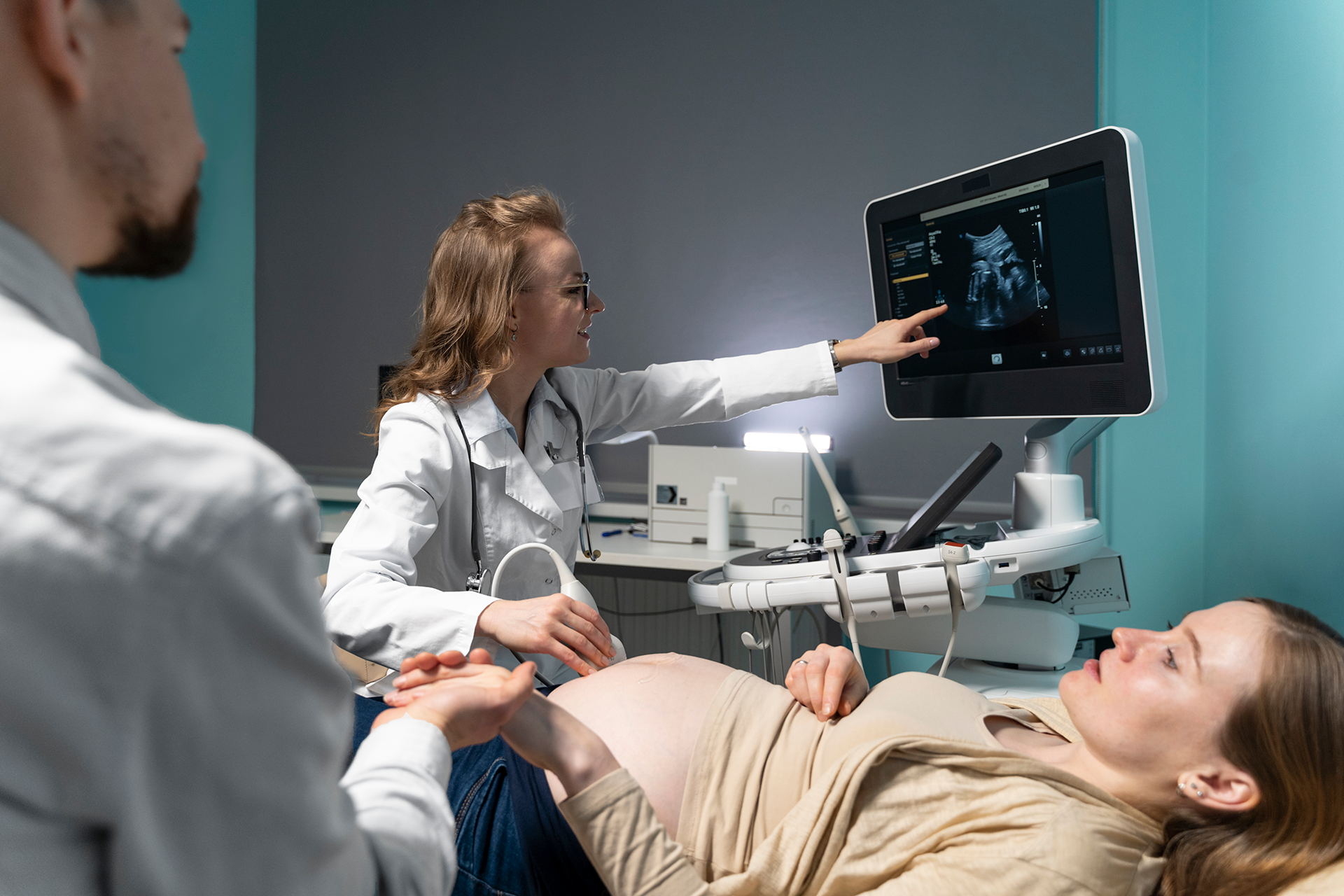
A pregnancy ultrasound, also known as an obstetric ultrasound, is a non‑invasive imaging test that uses high‑frequency sound waves to create real‑time images of the developing fetus, placenta, and maternal reproductive organs. Unlike X‑rays or CT scans, ultrasound does not use ionising radiation, making it the safest imaging modality for monitoring pregnancy.
A handheld device called a transducer is gently moved over the abdomen (or inserted vaginally in early pregnancy). The transducer emits sound waves that bounce off tissues and fluids; these echoes are then converted into moving images displayed on a monitor. This allows the radiologist to assess fetal growth, anatomy, position, and well‑being.
Why Are Ultrasound Scans Performed During Pregnancy?
Ultrasound serves multiple purposes throughout pregnancy, from confirming the pregnancy to preparing for delivery. Common reasons include:
- Confirming pregnancy and ruling out ectopic pregnancy
- Determining gestational age and estimating the due date
- Assessing fetal number (single, twins, or multiples)
- Evaluating fetal anatomy to detect structural abnormalities
- Monitoring fetal growth and amniotic fluid volume
- Placental location to identify conditions like placenta previa
- Guiding procedures such as amniocentesis
- Assessing fetal well‑being in high‑risk pregnancies
Types of Pregnancy Ultrasound Scans
First Trimester Scans (Weeks 6–13)
- Dating Scan (6–10 weeks): Confirms the pregnancy, measures the crown‑rump length to accurately date the pregnancy, detects the fetal heartbeat, and checks for multiple pregnancies.
- Nuchal Translucency (NT) Scan (11–13 weeks + 6 days): Measures the fluid collection at the back of the fetal neck. Combined with maternal blood tests, this forms the first‑trimester screening for chromosomal conditions such as Down syndrome.
Second Trimester Scan (Weeks 18–22)
- Anomaly Scan (Mid‑pregnancy Scan): A comprehensive examination of fetal anatomy. The radiologist systematically evaluates the brain, face, spine, heart, lungs, abdominal organs, limbs, and placenta. While most babies are developing normally, this scan can identify structural anomalies that may require further evaluation or planning.
Third Trimester Scans (Weeks 28–40)
- Growth and Well‑being Scans: Performed when there are concerns about fetal growth, low or excessive amniotic fluid, or in high‑risk pregnancies (e.g., maternal diabetes, hypertension). These scans assess fetal size, position, placental health, and blood flow in the umbilical artery using Doppler ultrasound.
Are Ultrasound Scans Safe?
Yes. Extensive research over decades has confirmed that diagnostic ultrasound is safe for both mother and baby when used appropriately. Ultrasound uses sound waves, not radiation, and there are no known harmful effects. At Alsafwa Radiology Center, we adhere to the ALARA principle (As Low As Reasonably Achievable), using the minimum acoustic output needed to obtain diagnostic information.
What to Expect During Your Ultrasound
Preparation
- For early pregnancy scans, you may be asked to have a full bladder to improve visibility of the uterus.
- For later scans, no special preparation is usually required. Wear comfortable, two‑piece clothing to allow easy access to your abdomen.
During the Scan
- You will lie on an examination table. A clear gel is applied to your abdomen to help the transducer make good contact.
- The radiologist or sonographer will move the transducer gently over your belly. You may feel slight pressure, but the procedure is painless.
- In early pregnancy, a transvaginal ultrasound (with a small, covered probe) may be performed for clearer images. This is entirely safe and often provides better detail in the first trimester.
- You will be able to see the images on a screen. The radiologist will explain what is being viewed, and you may receive printed or digital images of your baby.
Duration
Most scans take between 20 and 45 minutes, depending on the type and complexity. Anomaly scans may take longer because of the detailed assessment.
After the Scan
- You can resume normal activities immediately.
- A detailed report will be sent to your obstetrician, usually within 24 hours. Your doctor will discuss the results with you and recommend any necessary follow‑up.
The Role of the Radiologist in Prenatal Care
While ultrasound images are often the first “baby pictures” parents see, they also carry critical medical information. At Alsafwa Radiology Center, your scan is performed and interpreted by a specialist radiologist with advanced training in obstetric imaging. These physicians are skilled in recognising normal fetal anatomy and detecting subtle abnormalities that may require further evaluation. Their expertise ensures that your obstetrician receives an accurate, comprehensive report to guide your prenatal care.
Advanced Ultrasound Services at Alsafwa Radiology Center
- 3D/4D Ultrasound: Provides lifelike surface images of the baby’s face and movements. While primarily used for bonding and reassurance, it can also assist in evaluating certain facial anomalies.
- Doppler Ultrasound: Assesses blood flow in the umbilical cord, fetal vessels, and maternal uterine arteries—valuable in managing high‑risk pregnancies.
- Fetal Echocardiography: A dedicated scan of the fetal heart when a cardiac anomaly is suspected.
Our centre is equipped with modern ultrasound machines that deliver exceptional image clarity, ensuring that even the smallest details are visualised with confidence.
When Should You Have a Pregnancy Ultrasound?
The number and timing of ultrasounds vary based on individual circumstances. Most low‑risk pregnancies include:
- A dating scan in the first trimester
- An anomaly scan at 18–22 weeks
Additional scans may be recommended if you have a high‑risk condition, multiple gestation, or if the obstetrician needs to monitor specific parameters. Always follow the guidance of your healthcare provider.
Why Choose Alsafwa Radiology Center?
- Specialist Radiologists: Our team has extensive experience in obstetric imaging and works closely with your obstetrician.
- Advanced Technology: We use state‑of‑the‑art ultrasound equipment for clear, accurate imaging.
- Comfortable Environment: We understand that pregnancy is a special time. Our staff is trained to provide a calm, supportive experience.
- Convenient Location: Centrally located in Sharjah, we are easily accessible for expectant mothers across the emirate.
Conclusion
Pregnancy ultrasound scans are a cornerstone of modern prenatal care, offering invaluable insights into your baby’s health and development. At Alsafwa Radiology Center in Sharjah, we combine advanced imaging technology with compassionate expertise to support you throughout your pregnancy journey. Whether it’s your first glimpse of your baby or a detailed assessment later in pregnancy, you can trust that you are in caring, capable hands.
FAQ: Pregnancy Ultrasound
A: Yes. Decades of clinical use and research confirm that diagnostic ultrasound is safe for both mother and baby. It uses sound waves, not radiation, and is performed only when medically indicated.
A: For early pregnancy scans (first trimester), a full bladder helps lift the uterus into a better position for imaging. For mid‑pregnancy and later scans, a full bladder is usually not required unless specified.
A: First‑trimester ultrasound is highly accurate for determining gestational age, with an error margin of about 5–7 days. Later scans are less reliable for dating but are excellent for assessing growth.
A: Standard pregnancy ultrasounds are performed in 2D, which is sufficient for all medical assessments. 3D/4D imaging may be offered for additional visualisation or parental bonding, but it is not a routine medical requirement.
A: Ultrasound is an excellent screening tool, but it cannot detect every possible condition. Some abnormalities, especially genetic or functional issues, may not be visible on ultrasound. Your obstetrician will interpret the results in the context of your overall care.
A: Most scans take 20–45 minutes. The anomaly scan (mid‑pregnancy) is the longest because it involves a detailed review of all fetal structures.
A: Yes, we welcome one support person to accompany you during the scan. This is a special moment, and we are happy to share it with your loved ones.