Okay, let’s be honest. When your doctor says you need a scan, it’s easy for your mind to go blank after the acronym. CT, MRI, Ultrasound, X-Ray… they all sound like expensive, mysterious machines that see straight through you. (Which, to be fair, they kinda do).
But what’s the real difference? And why would your doctor pick one over the other? Think of it like this: doctors have a toolbox of imaging techniques, and each tool is perfect for a specific job. Let’s demystify the big four, so you can walk into your next appointment with a little more clarity and a lot less confusion.
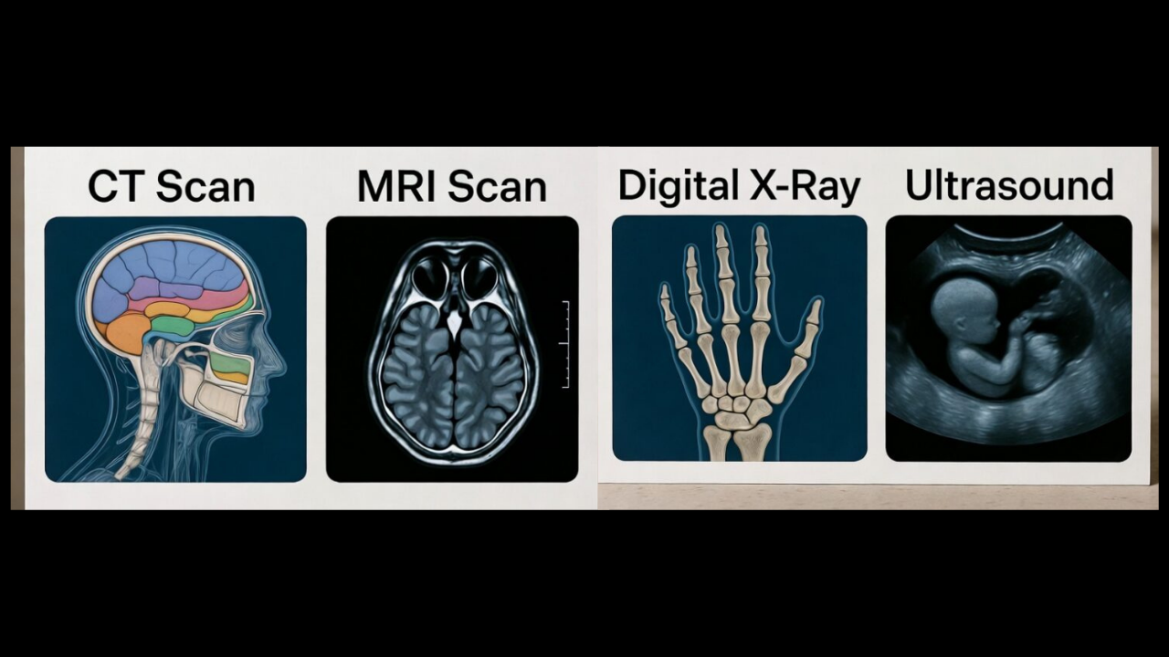
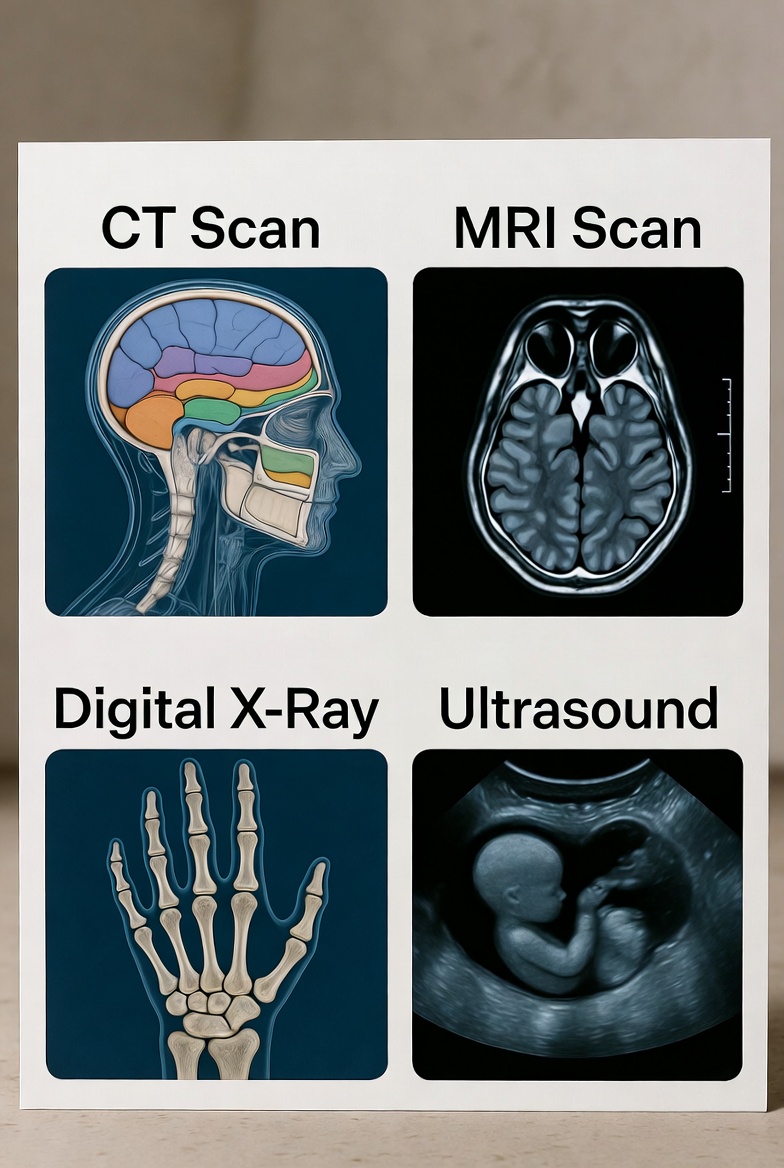
1. The Digital X-Ray: The Quick Snapshot
- How it works: Uses a very small, targeted dose of radiation to create a 2D picture, mostly of dense structures.
- The Quick Analogy: It’s like taking a black-and-white photo of your bones. Fast, clear, and great for capturing a single moment in time.
- Best for: The classics. Checking for broken bones (fractures), looking at teeth (dental X-rays), screening for pneumonia in the lungs, or getting a basic view of the chest or abdomen.
- The Patient Experience: It’s the fastest of the bunch. You stand, sit, or lie down, stay still for a second, and click—you’re often done in minutes. No noise, just a quick hold of your breath.
2. The CT Scan (Computed Tomography): The Detailed Cross-Section
- How it works: Takes a series of X-ray images from different angles and uses computer processing to create detailed cross-sectional “slices” of your body. It’s a 3D view built from many 2D pictures.
- The Quick Analogy: Imagine slicing a loaf of bread and looking at each individual slice. A CT scanner does that with your body, revealing what’s inside each “slice” in great detail.
- Best for: Emergencies (finding internal bleeding, a stroke, or a blood clot), detecting cancers, examining complex bone fractures, and getting a comprehensive look at organs like the lungs, liver, or kidneys. It’s fantastic for seeing a little bit of everything.
- The Patient Experience: You lie on a bed that moves through a large, doughnut-shaped ring. It’s painless but can be noisy. You might need a contrast dye (via drink or IV) to highlight certain areas. It’s relatively quick, often taking 10-20 minutes.
3. The MRI (Magnetic Resonance Imaging): The Soft Tissue Specialist
- How it works: Uses a powerful magnet and radio waves (no radiation) to create incredibly detailed images, especially of soft tissues. It looks at the composition of your tissues, not just their shape.
- The Quick Analogy: If a CT scan is great for the bones and structure of a house, an MRI is for examining the detailed wiring, plumbing, and drywall inside the walls.
- Best for: Imaging the brain, spinal cord, nerves, ligaments, tendons, and muscles. It’s the go-to for sports injuries (like torn ACLs or rotator cuffs), brain disorders, and detailed views of joints.
- The Patient Experience: You lie on a bed that slides into a long tube. The machine makes loud, repetitive knocking sounds (you’ll get earplugs or headphones). It requires you to stay very still for longer, often 30-45 minutes. Open MRI options are a fantastic alternative for those who feel anxious or are claustrophobic.
4. The Ultrasound: The Live, Dynamic View
- How it works: Uses high-frequency sound waves (like sonar) to create real-time moving images on a screen. No radiation is involved.
- The Quick Analogy: It’s like a submarine using sonar to map the ocean floor, but for your insides. It shows movement, like blood flowing or a baby kicking.
- Best for: Viewing developing babies during pregnancy, examining organs like the gallbladder, liver, uterus, ovaries, and bladder, and assessing blood flow in vessels (Doppler ultrasound). It’s interactive—the technician can see what’s happening as they move the probe.
- The Patient Experience: You lie down, a clear gel is applied to your skin, and a handheld probe (transducer) is moved over the area. It’s painless, radiation-free, and you might even get to watch the screen.
So, Which One is “Best”?
There isn’t a “best” one overall—only the best one for your specific situation. Your doctor chooses the tool based on what part of your body needs checking and what question they need answered. Sometimes, they might use two together to get the full picture.
Think of it this way:
- Broken arm? Start with an X-Ray.
- Unexplained headache or knee injury? An MRI might be the call.
- Car accident with potential internal injury? A CT is often the emergency workhorse.
- Expecting a baby or having gallbladder pain? Ultrasound is the classic choice.
The goal of all this amazing tech is the same: to help your medical team see clearly, so you can get the right diagnosis and the right care, faster.
FAQ: Your Diagnostic Imaging Questions, Answered
A: CT scans use a higher dose of radiation than a standard single X-ray because they take many X-ray images. However, the dose is carefully controlled and the medical benefit almost always outweighs the small risk. MRI and Ultrasound use no ionizing radiation.
A: Contrast agents are like “highlighter pens” for your scan. They help illuminate specific areas—like blood vessels on a CT or certain tissues on an MRI—making details much clearer and easier for the radiologist to interpret.
A: Absolutely. This is a very common concern. Many imaging centers now offer Open MRI machines, which are much less confining. Always tell your doctor and the imaging team about your anxiety beforehand—they can provide support, sometimes offer a mild sedative, or ensure you’re booked on an open machine.
A: It totally depends on the scan! You might be asked to fast (not eat or drink) for a few hours, avoid caffeine, or wear loose, metal-free clothing. The clinic will give you clear, specific instructions when you book your appointment.
A: A specialist doctor called a Radiologist analyzes the images in detail and creates a report for your referring doctor. For urgent cases, results can be very fast. For routine scans, it typically takes 24-48 hours for the full report to reach your doctor, who will then discuss the results with you.